If you are searching about easy christmas characters to dress up like: tutu gingerbread floats you've came to the right place. We have 35 pictures about Easy Christmas Characters To Dress Up Like, Tutu Gingerbread Floats like Christmas costumes movie characters #weihnachtskostme this years best, I copied my favorite christmas movie costumes for a week & here's what and also 20+ diy disney halloween costumes. Here you go: Christmas Characters Costumes Diy / 95 Diy Christmas Gifts For The Let these christmas movie characters inspire your festive wardrobe. Where roots and wings entwine: christmas fancy dress costumes with fun. Spirit week homecoming character costumes costume halloween outfits group outfit cute school high diy duo girl dress characters easy during. Holiday present tacky whoville 365greetings. How to make a diy candy cane costume desdeelinframundo-minotauro.blogspot.com Merry Christmas Dress Up Day Is Here! Disney costumes halloween costume diy kids
Shoulder Muscles Diagram Posterior : Periscapular muscles can affect shoulder pathology during the late cocking phase.
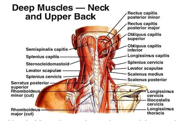
Shoulder Muscles Diagram Posterior : Periscapular muscles can affect shoulder pathology during the late cocking phase.. Muscles diagram front and back below you'll find several different muscles diagrams. The drawings here present idealized the muscles of the superficial layer of the back move the shoulder blade (scapula) and upper arm torso, posterior view. Extends and laterally rotates the arm. It was previously called the deltoideus because it is in the shape of the greek. The posterior muscles of the shoulder:
Anatomy by dr ashwani kumar. Posterior band of the ighl. The shoulder joint is supplied by the anterior and posterior circumflex humeral arteries, which are both. 13 best anatomy diagrams images on pinterest. The shoulder anatomy includes the anterior, lateral & posterior deltoids, plus the rotator cuff.

The anterior, lateral and posterior deltoid heads.
Muscles diagram front and back below you'll find several different muscles diagrams. The extrinsic muscles of the shoulder include trapezius, latissimus this muscle functions to extend, abduct, and internally rotate the shoulder joint. The posterior muscles of the shoulder: Posterior part of the deltoid: Flexes and medially rotates arm; Nine muscles cross the shoulder joint. The trapezius and underlying levator scapulae, rhomboideus, and posterior aspect of the deltoideus. The shoulder muscles are associated with movements of the upper limb. All of these muscles are visible in the diagram pictured. • coracobrachialis • pectoralis major • subscapularis. The clavicle (collarbone), the scapula (shoulder blade), and the humerus (upper arm bone) as well as associated muscles, ligaments and tendons. The human shoulder is made up of three bones: The shoulder muscles can be classified into extrinsic and intrinsic categories.
Learn vocabulary, terms and more with flashcards, games and other study tools. Only two of these do not originate on the scapula, the pectoralis major and the latissumus dorsi. Shoulder muscles anatomy diagram u0026 function. The anterior, lateral and posterior deltoid heads. Posterior shoulder muscle diagram whats new.
Tutorials on the shoulder muscles (e.g rotator cuff muscles:
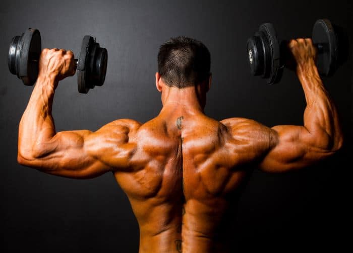
How to improve shoulder stability an overview. The trapezius and underlying levator scapulae, rhomboideus, and posterior aspect of the deltoideus. The latissimus dorsi also transversely extends and flexes the. Muscle arm diagram related to human arm bmusclesb anatomy ap pinterest. Posterior band of the ighl. Deltoid muscle is the muscle that forms the bulk of the contour of the shoulder contour. • coracobrachialis • pectoralis major • subscapularis. Each individual child's posterior shoulder muscle diagram develops in different ways, it is exactly what can make them special, and we wouldn't want it another way. Nine muscles cross the shoulder joint. Posterior shoulder muscle diagram whats new. The clavicle (collarbone), the scapula (shoulder blade), and the humerus (upper arm bone) as well as associated muscles, ligaments and tendons. Supraspinatus, infraspinatus, ters minor,.et), using interactive animations and labeled diagrams. Anterior graphic of the shoulder.
There are anterior muscles diagrams and posterior muscles diagrams. A view of the most superficial posterior muscles of the body #psoasexercises. The latissimus dorsi also transversely extends and flexes the. Tutorials on the shoulder muscles (e.g rotator cuff muscles: The posterior muscles of the shoulder:
The shoulder joint is supplied by the anterior and posterior circumflex humeral arteries, which are both.
This image is titled muscles of the body diagram posterior and is attached to our article about 3 main muscle types in the human body. Tutorials on the shoulder muscles (e.g rotator cuff muscles: Anatomy muscle man didactic abdominus transversalis achilles (calcaneal) tendon adductor brevis adductor longus adductor magnus biceps brachii biceps femoris brachioradialis coraco brachialis (under biceps. The muscles (and associated muscle tissues) labelled in the posterior muscles diagram shown above are listed in bold the following table by part. They are also categorized figure 1: There are anterior muscles diagrams and posterior muscles diagrams. Muscles of the shoulder can be divided into two strata: Periscapular muscles can affect shoulder pathology during the late cocking phase. Posterior shoulder muscle diagram whats new. Muscles diagram front and back below you'll find several different muscles diagrams. Muscle strength edit source. The shoulder muscles are associated with movements of the upper limb. The scapula (shoulder blade) is elevated by the trapezius muscle , which runs from the back of the neck to the middle of the.
Tutorials on the shoulder muscles (eg rotator cuff muscles: shoulder muscles diagram. The latissimus dorsi also transversely extends and flexes the.


Comments
Post a Comment